DIAGNOSIS YEAR
Select the diagnosis year first. J’Onco Mentor uses it to establish the standards cycle and to drive year-sensitive routing in this module.
Explore breast anatomy
Select a breast and quadrant. The utility suggests an ICD-O-3 primary-site code based on the selected region. For overlapping or multifocal patterns, use the documentation and SEER breast coding guidance rather than anatomy alone.
Selected breast & primary site
Select one or more breast regions.
Select one or more breast regions.
SEER 2026 coding reminder: when invasive tumor is documented in one subsite and in situ tumor in another, code the subsite containing the invasive tumor. Multiple tumors in at least two quadrants may lead to C50.9; a single tumor spanning subsites with origin unknown may lead to C50.8.
Cancer histology and ICD-O-3 coding
8500/2 — Ductal carcinoma in situ (DCIS)
METHOD OF DETECTION (MOD) — BREAST
NCDB/STORE data item 10110. Applies to breast primaries for diagnosis year 2026+.
—
Short explanation
Select a Method of Detection to display a short coding explanation.
Quick Coding Assistant
Choose the circumstance documented in the diagnostic imaging report.
Breast biomarkers & stage-related data
ER, PR, HER2, and Oncotype DX are maintained here as breast prognostic factors. Clinical and pathologic grade are entered in the AJCC Staging section. Residual Cancer Burden is a 2026 SSDI item; verify program requirements and source documentation before coding.
BREAST MULTIPLE PRIMARY RULES & CALCULATOR
Apply the Breast Solid Tumor Rules in order and stop at the first applicable rule.
Current Solid Tumor Rule updates matter: separate synchronous ductal and lobular tumors may be multiple primaries when the applicable histologies are on different rows of Table 3. Do not substitute receptor status for histology or primary determination.
AJCC Staging Schema
PATHOLOGIC STAGING CRITERIA — BREAST
Qualifying evidence: Lumpectomy, mastectomy or another qualifying excision generally supports pT. Sentinel-node biopsy or axillary dissection can support pN. Apply special criteria for in situ disease, microinvasion, and treatment before surgery.
pT / pN: Assign only categories supported by the actual pathological evidence and the site-specific AJCC rules. A diagnosis-confirming biopsy does not automatically establish a pathological primary tumor category.
pM: Pathologically proven distant metastasis may support pM1 where the chapter permits; AJCC does not use pM0.
Treatment sequence: Distinguish initial pathological classification (pTNM) from post-neoadjuvant classification (ypTNM). Confirm eligibility, histology, staging chapter and diagnosis-year edition before finalizing.
Source / verification: AJCC · SEER tools and manuals. Educational overview only; verify exact chapter-specific criteria in the licensed AJCC manual.
AJCC Breast: 8th Edition context for 2018–2026
Breast prognostic stage is not determined by T, N, and M alone. Biomarkers and grade can affect the prognostic stage group. This utility displays TNM components and supporting biomarker data rather than producing a definitive prognostic stage group.
Calculated from the selected AJCC 8th anatomic T, N and M categories. Prognostic stage may differ because grade, ER, PR and HER2 are also used.
Regional lymph nodes — breast
Use the realistic anatomy to review the major breast regional-node groups. Click node-group markers to identify documented involvement. Final cN/pN depends on nodal level, clinical versus pathologic assessment, positive-node count and deposit size.
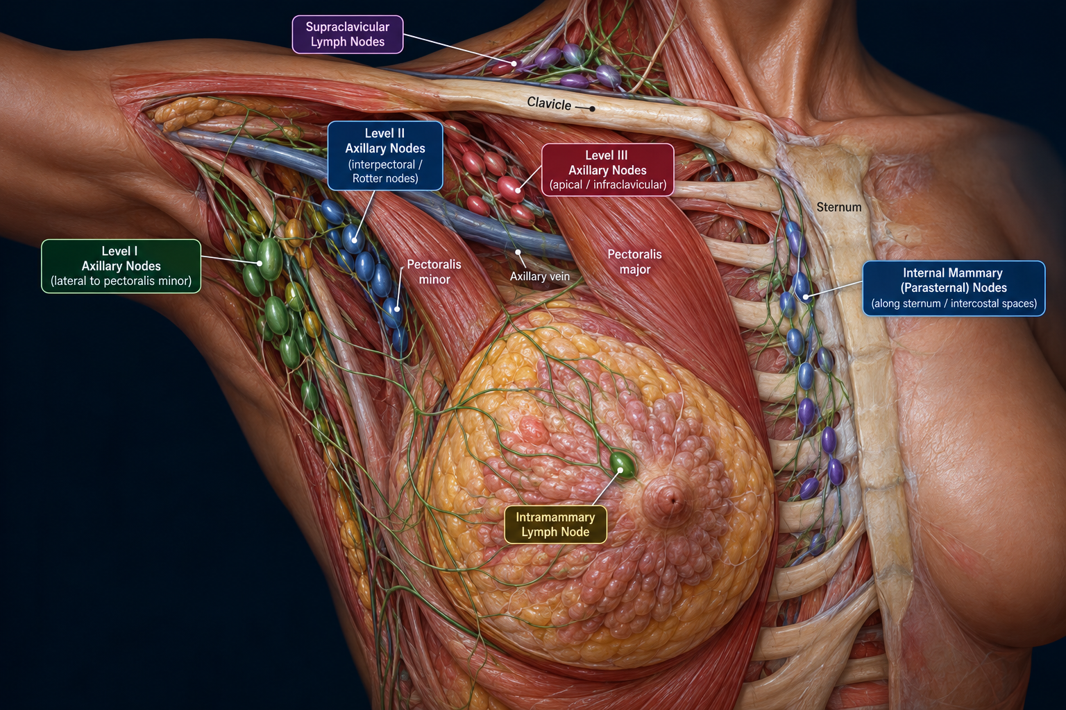
Anatomic context: Level I is lateral to pectoralis minor; Level II is behind/between its borders and includes interpectoral (Rotter) nodes; Level III is medial/apical/infraclavicular. Internal mammary nodes follow the parasternal intercostal spaces. Supraclavicular nodes are regional for breast staging.
None selected.
Select node groups above or enter the N category in the AJCC Staging section.
EOD — Breast
EOD Primary Tumor, EOD Regional Nodes, and EOD Mets are separate data items. Breast stage derivation additionally uses specific biomarker and grade data.
Use breast-specific extension and size rules from the applicable SEER*RSA version.
Use documented regional nodal groups, laterality, and extent. Do not infer an EOD code solely from the simplified node diagram.
Code documented distant metastatic disease using the schema-specific table.
SEER Summary Stage 2018
Noninvasive carcinoma confined to the epithelium with no invasion through the basement membrane. Includes in situ breast disease such as DCIS when no invasive component is present.
Invasive tumor confined to the breast without qualifying regional direct extension or regional lymph node involvement.
Select the applicable SEER Summary Stage 2018 Breast regional code.
Distant metastasis or distant lymph node involvement.
Insufficient information to assign a Summary Stage category.
Site-Specific Data Items (SSDI)
| Breast SSDI / Grade item | Why it matters |
|---|---|
| Grade Clinical / Pathological | Used for staging. |
| ER Summary | Used for staging. |
| PR Summary | Used for staging. |
| HER2 Summary | Used for staging. |
| Lymph Nodes Positive Axillary Level I-II | Used for staging. |
| Oncotype DX Recurrence Score | Stage-related breast data; applicability varies by year and context. |
| Residual Cancer Burden / RCB Class | 2026+ SSDI collection context. |
Breast Surgery Codes — 2026
B000
None
Breast reconstruction is recorded separately; review the operative report for the most definitive surgery.
Stage & Treatment
Treatment is individualized by stage, receptor status, HER2 status, patient factors, surgery feasibility, and multidisciplinary recommendations.
Educational only; use current treatment guidelines and physician documentation for clinical decision-making.
COMMON METASTATIC SITES — BREAST
Metastatic breast cancer most commonly involves the following distant sites.
Bone
A major and very common site of distant breast cancer involvement.
Lung
Common distant organ site.
Liver
Common distant organ site.
Brain
Recognized common site of metastatic breast cancer.
Sources
Official sources used throughout the Breast Primary page.