DIAGNOSIS YEAR
Select the diagnosis year first. J’Onco Mentor uses it to establish the standards cycle and to drive year-sensitive routing in this module.
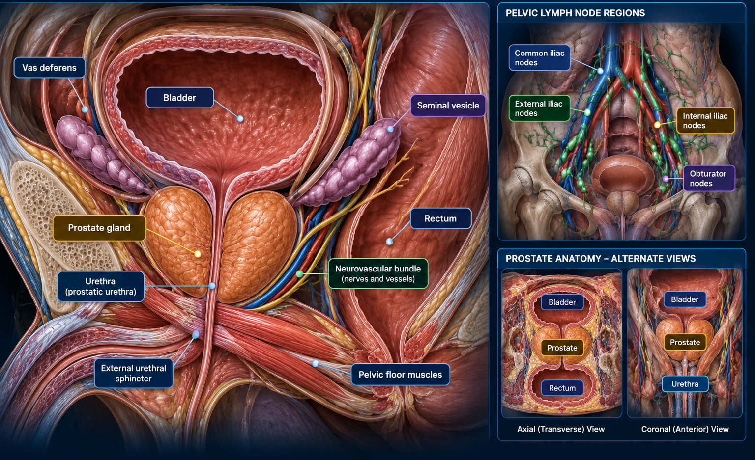
PROSTATE ANATOMY & PRIMARY SITE
Prostate gland
C61.9
Use C61.9 for a prostate gland primary. Transitional/urothelial carcinoma arising in the prostatic urethra is staged under the urethra schema rather than the prostate schema.
Bladder · prostate · prostatic urethra · seminal vesicles · neurovascular bundles · rectum · pelvic floor · external urethral sphincter
INTERACTIVE PROSTATE BIOPSY GUIDE
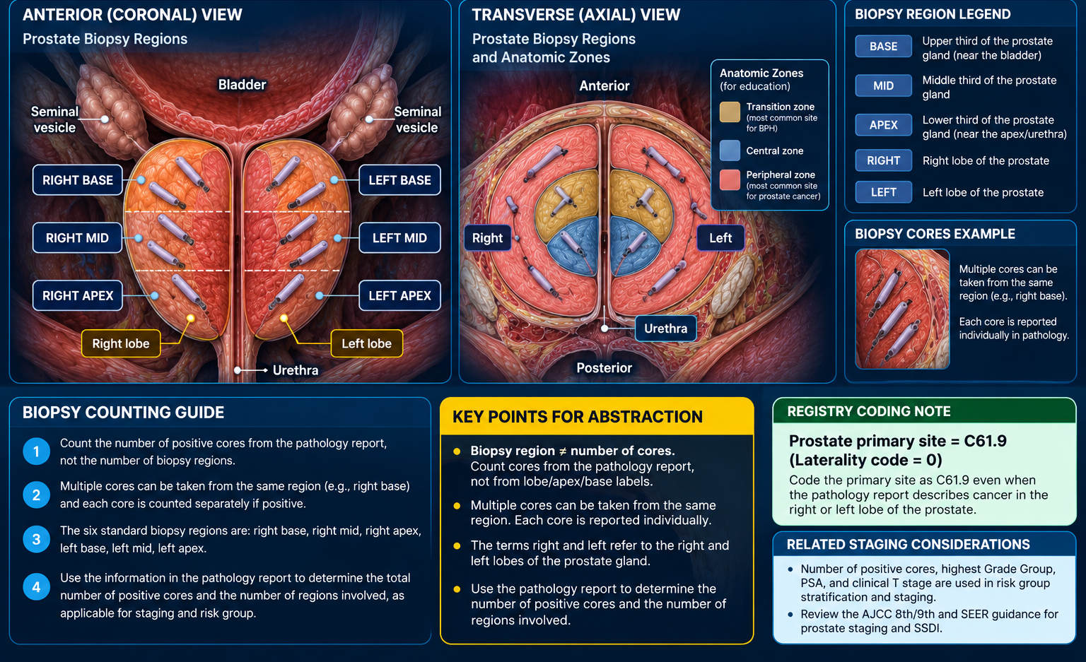
Click any biopsy region to mark it involved. Multiple positive cores may come from the same region, so region count is not the same as positive-core count.
CLINICAL T STAGING — MINIMUM EVIDENCE AND CRITERIA (PROSTATE)
Before assigning cT: Establish the documented clinical extent of the primary tumor using the applicable AJCC prostate chapter and pretreatment clinical assessment. Review the physician's clinical stage, digital rectal examination (DRE), relevant imaging, and biopsy findings. A biopsy confirms malignancy but the number or laterality of positive biopsy cores alone does not establish palpable tumor extent.
cT1: Clinically inapparent tumor that is not palpable. cT1c generally describes cancer identified by needle biopsy (for example, following an elevated PSA), when the tumor is not clinically apparent.
cT2: Tumor clinically apparent and confined within the prostate. The subcategory depends on clinically established lobe involvement: cT2a, one-half of one lobe or less; cT2b, more than one-half of one lobe but not both lobes; cT2c, both lobes.
cT3 / cT4: Documented extraprostatic extension or seminal vesicle invasion may support cT3 categories; fixation or invasion of qualifying adjacent structures may support cT4. Apply the AJCC chapter's exact definitions and evidence rules.
When evidence is insufficient: Do not infer a higher clinical T category from bilateral positive cores, a diagram selection, or PSA/Grade Group alone. Verify the physician's clinical findings and the diagnosis-year AJCC rules before assigning a category.
BIOPSY-ASSISTED cT CALCULATOR
AJCC STAGING
Educational reference: AJCC. Confirm chapter-specific staging criteria and edition in the licensed manual.
Prostate AJCC staging schema / TNM context
Use the T, N and M controls and site-specific factors below. Verify final AJCC ID, TNM edition and stage group in the approved AJCC/SEER staging system for the diagnosis year.
8th Edition
For 2026 prostate cases, stage grouping uses AJCC 8th Edition and incorporates T, N, M, PSA, and Grade Group.
The biopsy guide provides a suggested clinical T category and may synchronize it with the staging calculator. Verify the suggested category against physician-documented clinical extent and AJCC criteria before using it for staging.
PATHOLOGIC STAGING CRITERIA — PROSTATE
Qualifying evidence: Radical prostatectomy generally provides the tissue necessary to determine pathological primary tumor extent (pT). Prostate needle biopsy alone does not establish pT. Pathologically examined pelvic regional lymph nodes can support pN.
pT / pN: Assign only categories supported by the actual pathological evidence and the site-specific AJCC rules. A diagnosis-confirming biopsy does not automatically establish a pathological primary tumor category.
pM: Pathologically proven distant metastasis may support pM1 where the chapter permits; AJCC does not use pM0.
Treatment sequence: Distinguish initial pathological classification (pTNM) from post-neoadjuvant classification (ypTNM). Confirm eligibility, histology, staging chapter and diagnosis-year edition before finalizing.
Source / verification: AJCC · SEER tools and manuals. Educational overview only; verify exact chapter-specific criteria in the licensed AJCC manual.
PROSTATE PATHOLOGIC STAGE CALCULATOR — pTNM / ypTNM
Enter only pathologically documented categories. This evidence-based worksheet displays the entered classification; it does not infer a final AJCC prognostic stage group. Confirm eligibility, treatment sequence, and stage grouping in the applicable AJCC manual.
A manual value overrides the quick-select dropdown.
INTERACTIVE PROSTATE LYMPH NODES & CLINICAL N CALCULATOR
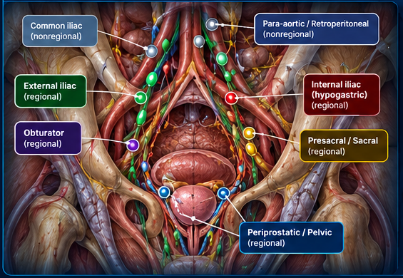
Click any highlighted nodal group to toggle it. Regional pelvic nodes contribute to N category; nonregional nodes contribute to M1a.
cN0
No positive regional pelvic nodes identified.
cM0
No nonregional lymph node metastasis identified.
000
No regional lymph node involvement.
External iliac · Internal iliac / hypogastric · Obturator · Periprostatic / pelvic · Presacral / sacral
Common iliac · Para-aortic / retroperitoneal
ICD-O-3 HISTOLOGY
SEER 2027 — PROSTATE MULTIPLE PRIMARY RULES
INTERACTIVE PROSTATE MULTIPLE PRIMARY REVIEW
—
Select the pattern and calculate.
PROSTATE-SPECIFIC RULES
M3 — Acinar adenocarcinoma: Acinar adenocarcinoma of the prostate (8140) is always a single primary.
Report only one acinar/adenocarcinoma of prostate per patient lifetime. This also applies to qualifying subtype/variant forms listed with acinar adenocarcinoma.
M4 — Subsequent small cell carcinoma: Abstract multiple primaries when a subsequent small cell carcinoma of prostate occurs more than one year after acinar adenocarcinoma and/or an acinar subtype/variant.
OTHER-SITES RULES THAT CAN MATTER
M19 — Distinct histologies: Separate/non-contiguous tumors on different rows of the current Other Sites histology table are multiple primaries; timing is irrelevant.
SEER specifically confirms that separate acinar adenocarcinoma and small cell carcinoma diagnosed less than one year apart can still be separate primaries under M19 when M4 does not apply.
M20 — In situ then invasive: A separate invasive tumor occurring more than 60 days after an in situ tumor is abstracted as a new primary.
SURGERY OF PRIMARY SITE — 2026
—
No surgery selected.
CODING NOTEDo not code orchiectomy in Surgery of Primary Site. For prostate primaries, orchiectomy is coded in Hematologic Transplant and Endocrine Procedures (NAACCR #3250).
PSA BY AGE — REFERENCE RANGES
| Age | Common age-specific upper reference |
|---|---|
| 40–49 | ≈ 2.5 ng/mL |
| 50–59 | ≈ 3.5 ng/mL |
| 60–69 | ≈ 4.5 ng/mL |
| 70–79 | ≈ 5.5 ng/mL |
These are age-specific reference limits, not a universal “average PSA.” PSA interpretation varies by age, race, prostate size, medications, infection/inflammation, laboratory reference range, and clinical context.
For cancer registry abstraction, code the qualifying pretreatment total serum PSA according to the SEER SSDI instructions. Do not substitute free PSA or precursor PSA.
AMERICAN CANCER SOCIETY — UPDATED PROSTATE CANCER SCREENING GUIDELINES (2026)
The American Cancer Society has updated its guidelines to recommend routine prostate-specific antigen screening every two years for 50- to 69-year-old men at average risk.
The updated guideline recommends earlier and more frequent screening for men at higher risk and adds a more structured pathway for confirming and evaluating an elevated PSA.
Average risk: PSA-based screening every 2 years from ages 50–69.
High risk: Black men and non-Black men with a strong family history should undergo annual PSA-based screening from ages 45–69.
Family-history nuance: Non-Black men with one first-degree relative diagnosed at age 65 or older should be screened every 2 years from ages 45–69.
Ages 45–69: PSA ≥ 3.0 ng/mL is considered elevated under the updated ACS screening pathway.
Ages 70–74: PSA ≥ 6.5 ng/mL is considered elevated.
Repeat an elevated PSA between 2 weeks and 3 months. If the elevation is confirmed, offer MRI and/or a validated biomarker to help guide the biopsy decision.
EXTENT OF DISEASE (EOD)
Code prostate extension using the 2026 SEER EOD prostate schema.
Code regional pelvic nodal involvement separately from distant nodes.
Capture distant metastases including bone, distant nodes, and other organs as defined in the schema.
SEER SUMMARY STAGE 2018
Localized · Regional · Distant · Unknown. Apply the current prostate-specific Summary Stage 2018 rules rather than inferring directly from AJCC stage group.
AJCC, EOD, and Summary Stage are separate staging systems. Do not substitute one system's codes for another.
PROSTATE SSDI / STAGING FACTORS
Pretreatment prostate-specific antigen value is needed for AJCC stage grouping.
Grade Group 1–5 based on clinical-time-frame Gleason findings.
Pathologic Grade Group when a qualifying pathologic specimen/time frame exists.
1 — Gleason ≤6 · 2 — Gleason 3+4=7 · 3 — Gleason 4+3=7 · 4 — Gleason 8 · 5 — Gleason 9–10
COMMON METASTATIC SITES — PROSTATE
Bone is a particularly important metastatic site in prostate cancer. Other recognized distant sites include liver, lung, adrenal gland, and distant lymph nodes.
Bone
Bone metastases are common in prostate cancer and are specifically addressed in the prostate EOD Mets schema.
Distant lymph nodes
Examples include aortic, common iliac, inguinal, and retroperitoneal nodes when classified as distant by schema.
Liver
Recognized distant organ site.
Lung
Recognized distant organ site.
Adrenal gland
Listed by NCI among main sites of prostate cancer metastasis.