DIAGNOSIS YEAR
Select the diagnosis year first. J’Onco Mentor uses it to establish the standards cycle and to drive year-sensitive routing in this module.
INTERACTIVE CNS PRIMARY-SITE SELECTOR
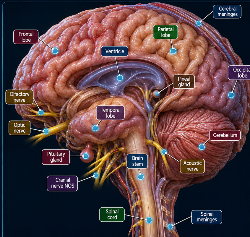
Select a cyan marker on the realistic CNS atlas to drive the primary-site code and laterality logic. Click the active marker again to deselect it.
Expanded CNS histology selector in the Primary Site box. It includes malignant and non-malignant CNS choices used in current SEER/ICD-O-oriented workflows and stays synchronized with the Histology section. Rare terms not listed should still be verified in current ICD-O-3.2 / Solid Tumor Rules.
Selecting an additional site immediately adds it to the anatomy selection and recalculates the primary-site result.
- No regions selected.
REPORTABILITY & BEHAVIOR
Malignant CNS
Use the current Malignant CNS and Peripheral Nerves Solid Tumor Rules for applicable reportable sites/histologies.
Benign / Borderline CNS
Intracranial and intraspinal benign/borderline tumors can be reportable. For spinal tumors, intradural location is important; extracranial/extradural nerve tumors may not meet CNS reportability rules.
Intracranial glands
Pituitary, craniopharyngeal duct, and pineal gland are included in the CNS Solid Tumor Rules site groups when applicable.
CNS HISTOLOGY / TUMOR FAMILY
Integrated CNS diagnosis
Modern CNS diagnoses may combine morphology with molecular findings. Use the final documented integrated diagnosis and the current ICD-O-3.2 / SEER Solid Tumor Rules when assigning the definitive histology and behavior.
The same expanded histology menu is available in the Primary Site selector above. Rare or newly introduced terms should be verified in current ICD-O-3.2 / Solid Tumor Rules.
Diffuse astrocytic tumors
Includes astrocytoma and glioblastoma families. Molecular status can be integral to the final WHO diagnosis.
Oligodendroglial tumors
Oligodendroglioma diagnosis incorporates IDH mutation and 1p/19q co-deletion status.
Ependymal / embryonal
Includes ependymal tumors, medulloblastoma, and other embryonal families.
MULTIPLE PRIMARY RULES — CNS
Use the correct CNS rule set
Malignant CNS and Non-Malignant CNS have distinct site-group instructions and tables. Do not apply the Other Sites rules to CNS tumors.
Site + histology relationships matter
CNS multiple-primary decisions can depend on whether tumors are on the same or different rows of the current Solid Tumor Rules tables, as well as timing and transformation/progression rules.
MOLECULAR MARKERS / SSDI
Brain Molecular Markers
Captures clinically important molecularly defined brain tumor subtypes that may not be distinguishable by the base ICD-O-3 code alone.
MGMT methylation
Used for applicable brain histologies. Record the documented methylation result according to the current schema.
Integrated diagnosis
WHO CNS diagnoses increasingly combine histology with molecular findings. Do not substitute a molecular marker result for the documented final integrated diagnosis.
WHO CNS GRADE
Use the documented grade and current Grade Manual rules. Numeric WHO grades take priority when available.
Grade data items
Grade may be collected as Clinical, Pathological, Post-Therapy Clinical, or Post-Therapy Pathological depending on the case and treatment sequence.
AJCC STAGING SCHEMA / TNM ENTRY
PATHOLOGIC STAGING CRITERIA — CNS / BRAIN
Qualifying evidence: Most primary CNS tumors do not use conventional AJCC pTNM. Use the applicable WHO CNS tumor classification, histology, molecular findings and grade. Do not generate pT or pN categories where no AJCC TNM schema applies.
pT / pN: Assign only categories supported by the actual pathological evidence and the site-specific AJCC rules. A diagnosis-confirming biopsy does not automatically establish a pathological primary tumor category.
pM: Pathologically proven distant metastasis may support pM1 where the chapter permits; AJCC does not use pM0.
Treatment sequence: Distinguish initial pathological classification (pTNM) from post-neoadjuvant classification (ypTNM). Confirm eligibility, histology, staging chapter and diagnosis-year edition before finalizing.
Source / verification: AJCC · SEER tools and manuals. Educational overview only; verify exact chapter-specific criteria in the licensed AJCC manual.
TNM CAPTURE
| T | — |
|---|---|
| N | — |
| M | — |
—
Use the CNS STAGING section for the actual SEER schema and Summary Stage workflow.
CNS STAGING
SCHEMA IDENTIFICATION
STAGING METHOD
The applicable staging method will appear after schema identification.
EOD Primary Tumor
Use the selected schema to code confinement, extension within the CNS, crossing the midline when specifically documented, and extension to adjacent structures.
EOD Regional Nodes
Regional lymph-node staging is generally not applicable to primary CNS-schema tumors. Follow the schema-specific instructions.
EOD Mets / Dissemination
Captures qualifying discontinuous spread, including CNS/CSF dissemination such as leptomeningeal or drop metastases and extra-neural distant disease when defined by the schema.
TREATMENT / SURGERY REFERENCE
Resection
Document extent of surgery from operative and pathology reports. Gross total, subtotal, and biopsy-only procedures should not be inferred from imaging language alone.
Radiation
Capture modality, target, and course according to STORE/NAACCR rules; CNS treatment may include focal radiation, stereotactic techniques, or craniospinal fields depending on disease.
Systemic therapy
Therapy varies substantially by histology and molecular profile. This section is educational and should not be used as a treatment recommendation engine.
COMMON SPREAD / DISSEMINATION — PRIMARY CNS
Primary CNS tumors more commonly disseminate within the CNS/CSF pathways than to extracranial organs. Patterns are highly histology-dependent.
Leptomeninges / CSF
Leptomeningeal dissemination and positive CSF can represent distant CNS spread depending on the schema context.
Spinal canal / “drop” metastases
Seen with selected CNS histologies and captured in EOD Mets when discontinuous spread is documented.
Other CNS sites
Discontiguous intracranial or intraspinal spread may occur depending on histology.
Extra-neural metastasis
Possible but uncommon for primary CNS tumors; code only when documented.
SOURCES
Primary site, reportability, coding guidanceSEER 2026 — Brain/CNS Benign & Borderline
Reportability and CNS site guidanceSEER 2026 Solid Tumor Rules
Malignant CNS and Non-Malignant CNS rule setsSEER*RSA EOD v3.3 — NAACCR 2026
Brain and CNS Other schemas, grade, SSDIsSEER*RSA — Brain V9
Brain schema for applicable 2023+ casesSEER*RSA — CNS Other V9
Spinal cord, meninges, cranial nerve and other CNS routingSEER*RSA — Medulloblastoma V9
Separate 2023+ schema for eligible medulloblastoma/embryonal histologiesSEER Training — CNS
Anatomy, pathology, grade, molecular testingSEER*RSA — Intracranial Gland V9
Schema selection and staging applicabilityNCI — AJCC Soft Tissue Sarcoma
AJCC 8 head & neck T definitions and stage-group limitation