DIAGNOSIS YEAR
Select the diagnosis year first. J’Onco Mentor uses it to establish the standards cycle and to drive year-sensitive routing in this module.
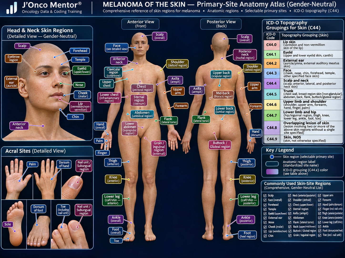
INTERACTIVE CUTANEOUS MELANOMA ANATOMY / PRIMARY-SITE MAP
Click one or more highlighted points on the anatomy atlas or use the site buttons. Multiple sites can remain selected at the same time. Click any selected hotspot, quick chip, or site button again to deselect it. The ICD-O C44.x field updates from the current selection set, and a clear-all button removes every selection at once.
SELECTED ANATOMIC REGIONS
The atlas identifies anatomic regions. ICD-O topography is assigned at the C44.x grouping level; detailed labels such as abdomen, flank, buttock, hand, sole, and nail unit map into their applicable C44.x site group.
Cutaneous Melanoma Solid Tumor Rules apply to C44.0–C44.9 with histologies 8720–8780. Melanoma originating in mucosal, ocular, or other non-skin sites uses a different rule/schema family.
SHOW TEXT-ONLY SITE BUTTONS
HISTOLOGY
REPORTABILITY / HISTOLOGY NOTES
For diagnoses beginning in 2021, SEER melanoma guidance treats early/evolving melanoma terminology as reportable when it represents in situ or invasive melanoma.
Use the current Cutaneous Melanoma Histology Rules to resolve NOS versus subtype/variant combinations. Do not create additional primaries solely because biomarkers differ.
Rare melanoma histologies may require the current ICD-O update and/or SEER inquiry if not found in the melanoma table.
SEER 2027 — CUTANEOUS MELANOMA MULTIPLE PRIMARY RULES & CALCULATOR
—
Select the case pattern and calculate.
ORDERED RULE SUMMARY
M1: Unknown whether single or multiple after all sources exhausted → single primary.
M2: One melanoma → single primary.
M3: Separate non-contiguous melanomas with different ICD-O site codes → multiple primaries.
M4: Separate non-contiguous melanomas with different lateralities → multiple primaries when laterality applies.
CONTINUED RULES
M5: Different melanoma subtype/variants in Table 2 Column 2 → multiple primaries.
M6: Synchronous tumors on the same Table 2 row and same site code → single primary.
M7: Melanomas diagnosed more than 60 days apart → multiple primaries.
M8: If none of the prior rules apply → single primary.
REGIONAL LYMPH NODES / AJCC N
Use the interactive melanoma lymph-node atlas to identify a regional basin. Select one region at a time; click the selected hotspot or basin button again to deselect it, or use Clear All.
MELANOMA LYMPH-NODE REGION ATLAS
Same anterior/posterior anatomy used in the melanoma primary-site atlas. Cyan markers are selectable lymph-node regions.
REGIONAL NODE BASINS
Regional basins depend on the skin primary site and lymphatic drainage pattern. Common basins include cervical, axillary, epitrochlear, inguinal/femoral, and popliteal nodes.
Sentinel lymph-node findings can change pathologic N and therefore pathologic stage grouping. Do not classify a distant nodal basin as regional without checking the applicable melanoma schema/summary-stage guidance.
AJCC STAGING
PATHOLOGIC STAGING CRITERIA — CUTANEOUS MELANOMA
Qualifying evidence: Pathological evaluation of the primary melanoma establishes Breslow thickness and ulceration for pT; biopsy material may suffice when adequate. Sentinel lymph node biopsy or regional node dissection can establish pN. Do not require wide excision for every pathological T determination.
pT / pN: Assign only categories supported by the actual pathological evidence and the site-specific AJCC rules. A diagnosis-confirming biopsy does not automatically establish a pathological primary tumor category.
pM: Pathologically proven distant metastasis may support pM1 where the chapter permits; AJCC does not use pM0.
Treatment sequence: Distinguish initial pathological classification (pTNM) from post-neoadjuvant classification (ypTNM). Confirm eligibility, histology, staging chapter and diagnosis-year edition before finalizing.
Source / verification: AJCC · SEER tools and manuals. Educational overview only; verify exact chapter-specific criteria in the licensed AJCC manual.
Melanoma of Skin AJCC staging schema / TNM context
Use the T, N and M controls and site-specific factors below. Verify final AJCC ID, TNM edition and stage group in the approved AJCC/SEER staging system for the diagnosis year.
BRESLOW-ASSISTED T CALCULATOR
DISTANT METASTASIS
AJCC 8 uses M1a–M1d by metastatic site and incorporates LDH status as an M-category suffix/detail. Any M1 category is Stage IV.
CLINICAL TNM
PATHOLOGIC TNM
EOD — MELANOMA SKIN (NAACCR 2026)
EOD PRIMARY TUMOR
Use the current Melanoma Skin EOD schema to code the primary tumor extent. Breslow thickness and ulceration are captured separately as SSDIs and should not be substituted for the EOD primary-tumor code.
EOD REGIONAL NODES
Code regional nodal involvement using the melanoma-specific EOD regional-node table, including applicable satellite/in-transit/microsatellite patterns.
EOD METS
Code distant metastatic disease using the current EOD Mets table; distant skin/subcutaneous tissue, lung, non-CNS visceral sites, and CNS involvement are clinically important metastatic patterns.
SUMMARY STAGE 2018
0 — IN SITU
Melanoma confined to the epidermis / in situ.
1 — LOCALIZED
Invasive melanoma confined to the skin primary site without regional or distant spread.
REGIONAL
Regional nodal involvement and/or melanoma-specific regional satellite, microsatellite, or in-transit disease is coded according to the current Summary Stage/EOD schema.
7 — DISTANT
Distant skin/subcutaneous tissue, distant lymph nodes, lung, CNS, or other distant visceral involvement.
9 — UNKNOWN
Insufficient information to determine extent.
SSDI / GRADE
MELANOMA-SPECIFIC SSDIs
- Breslow Tumor Thickness — NAACCR #3817
- Ulceration — NAACCR #3936
- Mitotic Rate Melanoma — NAACCR #3893
- LDH Lab Value — NAACCR #3932
- LDH Upper Limits of Normal — NAACCR #3870
- LDH Level — NAACCR #3869
- Clinical Margin Width — NAACCR #3961
GRADE / NEOADJUVANT ITEMS
The current 2026 melanoma schema also includes Grade Clinical, Grade Pathological, post-therapy grade fields, Neoadjuvant Therapy, Clinical Response, and Treatment Effect where required by the reporting standard.
Use the current SEER*RSA schema for exact allowable values and registrar notes.
2026 SKIN SURGERY CODES
B000
None / autopsy only
CODING REMINDERS
SEER instructs registrars to code the procedure from the operative description using the priority order of operative report, physician statement, pathology procedure description, then pathology results.
Do not assign the surgery code from margin status alone.
COMMON METASTATIC SITES — MELANOMA OF SKIN
Melanoma may spread through lymphatic and hematogenous routes and can involve many organs.
Regional lymph nodes
SEER Training identifies regional lymph nodes as the most common site of melanoma metastasis.
Skin / soft tissue
Common metastatic involvement, including in-transit/satellite patterns when applicable to staging.
Lung
Common hematogenous distant site.
Liver
Common hematogenous distant site.
Brain / bone
Both are recognized common distant metastatic sites.