DIAGNOSIS YEAR
Select the diagnosis year first. J’Onco Mentor uses it to establish the standards cycle and to drive year-sensitive routing in this module.
INTERACTIVE KIDNEY PRIMARY-SITE SELECTOR
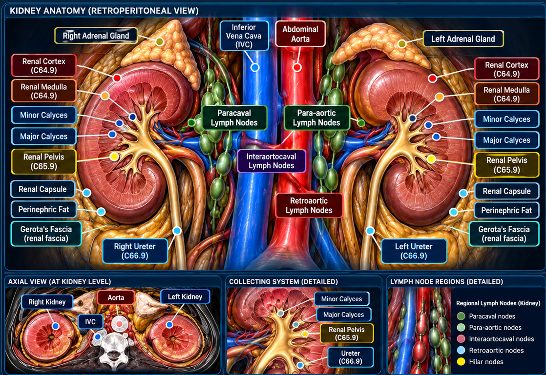
Select the documented site or sites of origin directly on the updated bilateral kidney diagram. The tool will calculate primary-site interpretation and laterality. Kidney parenchyma uses C64.9; renal pelvis uses C65.9; ureter uses C66.9.
—
Laterality is read directly from the selected diagram hotspot: anatomical right kidney = Right; anatomical left kidney = Left.
Select one or more anatomic structures.
C64.9 — Kidney (renal parenchyma)
C65.9 — Renal pelvis / calyceal collecting system
C66.9 — Ureter
KIDNEY HISTOLOGY & SCHEMA ROUTING
KIDNEY MULTIPLE PRIMARY RULES & CALCULATOR
INTERACTIVE KIDNEY MULTIPLE PRIMARY REVIEW
—
Select the case pattern and calculate.
KEY KIDNEY RULES
M1: If it remains unknown whether there is one tumor or multiple tumors after all information sources are exhausted, abstract a single primary.
M2: A single tumor is a single primary.
M3: Multiple tumors in sites whose ICD-O site codes differ at the second, third, and/or fourth character are multiple primaries.
M4: Bilateral nephroblastomas (Wilms tumors) are a single primary regardless of timing.
M5: Tumors in both the right and left kidney are multiple primaries unless pathology proves the contralateral tumor(s) are metastatic from the other kidney.
IMPORTANT KIDNEY NOTES
- Kidney cortex, medulla, capsule, sinus/hilum and renal parenchyma all remain C64.9; they are not separate C64 subsites.
- Renal pelvis C65.9 and ureter C66.9 are different primary-site codes and use a different urinary-site rule set.
- Biomarker differences do not by themselves create multiple primaries.
- If multiple tumors remain in the same unilateral kidney and no early rule resolves the case, continue through the current Kidney rules.
INTERACTIVE KIDNEY REGIONAL LYMPH NODES & cN CALCULATOR
cN0
No involved regional lymph node groups selected → cN0.
Use Auto for diagram-driven staging, or manually override the Clinical N category when it is documented elsewhere.
AJCC STAGING
PATHOLOGIC STAGING CRITERIA — KIDNEY
Qualifying evidence: Partial or radical nephrectomy generally supports pT assessment of renal tumor size and extension. Regional lymph node pathology supports pN when available. Verify the applicable renal primary and histology chapter.
pT / pN: Assign only categories supported by the actual pathological evidence and the site-specific AJCC rules. A diagnosis-confirming biopsy does not automatically establish a pathological primary tumor category.
pM: Pathologically proven distant metastasis may support pM1 where the chapter permits; AJCC does not use pM0.
Treatment sequence: Distinguish initial pathological classification (pTNM) from post-neoadjuvant classification (ypTNM). Confirm eligibility, histology, staging chapter and diagnosis-year edition before finalizing.
Source / verification: AJCC · SEER tools and manuals. Educational overview only; verify exact chapter-specific criteria in the licensed AJCC manual.
Kidney AJCC staging schema / TNM context
Use the T, N and M controls and site-specific factors below. Verify final AJCC ID, TNM edition and stage group in the approved AJCC/SEER staging system for the diagnosis year.
CLINICAL T / N / M
Use the clinical/pretreatment size documented in the diagnostic workup. T1/T2 require a known size when confined to the kidney.
cTX
cN0
cM0
—
PATHOLOGIC T / N / M
Use the resection/pathology size when pathologic classification criteria are met.
—
—
—
EXTENT OF DISEASE (EOD)
Confined to kidney / renal capsule / cortex-medulla.
Regional extension including renal vein/major vessels, renal sinus/perirenal fat, IVC, or extension beyond Gerota fascia depending on exact structure.
Further contiguous extension such as aorta, liver, ribs, spleen, stomach.
SUMMARY STAGE 2018
Confined to kidney, including capsule/cortex/medulla.
Direct extension only / regional nodes only / both direct extension and regional nodes.
Distant site(s) or distant lymph nodes.
SSDI / GRADE
Preferred kidney grading uses Grade 1–4 when documented. Assign the highest grade from the primary tumor.
Invasion Beyond Capsule · Ipsilateral Adrenal Gland Involvement · Major Vein Involvement · Sarcomatoid Features.
SURGERY OF PRIMARY SITE — 2026
—
No surgery selected.
COMMON METASTATIC SITES — KIDNEY
Renal cell carcinoma has a broad hematogenous metastatic pattern.
Lung
Common hematogenous site of spread from kidney parenchymal cancers.
Bone
Common distant site.
Liver
Common distant organ site.
Brain
Recognized metastatic site, especially for kidney parenchymal primaries.
Adrenal gland
NCI lists adrenal involvement among main sites of kidney cancer metastasis.